
Understanding Spondylolisthesis
Spondylolisthesis
Spondylolisthesis is a condition in which one vertebra slips forward over the vertebra below, causing back pain and possible neurological problems.
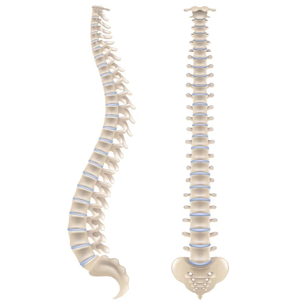
What is Spondylolisthesis?
Spondylolisthesis is the progressive slippage of one vertebra over the underlying vertebra. It can occur due to a bone fracture of one of the structures that connect the vertebrae (isthmic lysis), causing symptoms in young adults (15-45 years), or it can be the result of a form of osteoarthritis that deforms the joints between the vertebrae, often accompanied by narrowing of the lumbar canal, usually after age 60. The latter case is addressed in the section on lumbar spinal stenosis.
Evolution of Spondylolisthesis

SYMPTOMS
Isthmic lysis can, in rare cases, be diagnosed at the moment it occurs, usually in childhood or adolescence, manifesting as intense low back pain. In most other cases, the fracture occurs gradually and silently, being known as a fatigue fracture. In many patients, spondylolisthesis is diagnosed later, through an X-ray requested to evaluate low back pain that does not respond well to analgesic treatment. Furthermore, sciatica may appear, often limited to the buttocks or the back of the thighs. In cases of significant slippage of the vertebra, there may be visible deformity in the lower back and hip. Rarely, signs of paralysis in the legs may appear.
TESTS
A simple X-ray of the lumbar spine is, in most cases, sufficient to diagnose spondylolisthesis. Computed tomography (lumbar CT scan) is essential to evaluate fractures and bony contours in general. Magnetic resonance imaging (MRI) plays an important role in assessing the state of the disc between the slipping vertebrae, as well as the adjacent discs.
Treatment Options
Treatment Options for Spondylolisthesis
The initial treatment for lower back pain is always medical. It can be managed by the attending physician, often in collaboration with a rheumatologist, a physiatrist, or a pain specialist. This treatment includes rest during flare-ups, analgesic and anti-inflammatory medications prescribed on a progressive scale, physiotherapy, and, in some cases, infiltrations or even immobilisation with a brace. Most back pain responds well to these treatments, even if the flare-ups do not disappear completely. A more in-depth evaluation by a spinal surgeon is recommended when the pain does not respond adequately to these treatments and the symptoms, persistent for at least 6 to 12 months, cause significant limitations in daily life.
Principles of Surgical Treatment
Critérios para Cirurgia da Espondilolistese
Surgery for chronic low back pain caused by spondylolisthesis should only be considered after the failure of a long and complete medical treatment. The principle of surgery is to perform a fusion (arthrodesis) between the two unstable vertebrae, since the natural means of uniting these vertebrae are no longer sufficient. This bone fusion must be done after repositioning the slipped vertebra into the correct position, to prevent the development of a deformity that could cause premature degeneration of the adjacent discs.

Surgical Procedure
Lumbar Fusion
Surgery for spondylolisthesis has two main goals: to correct the spinal deformity (spondylolisthesis reduction) and to fuse the vertebrae (arthrodesis). It can be performed using different techniques, which do not always have the same objectives or results.
The first intervention can be performed through the abdomen, minimizing tension on the nerve roots located behind the disc. It is a minimally invasive technique, with an incision of 5 to 7 cm in slender patients, allowing the removal of the disc and the placement of an implant (fusion cage) fixed to the two vertebrae. In this step, the spondylolisthesis is corrected and the spine is rebalanced, preventing problems in the adjacent discs in the future. The second intervention is simpler, also minimally invasive or percutaneous, and consists of placing metallic fixation implants (osteosynthesis) from the posterior side, to ensure the best conditions for vertebral fusion. The positioning of the rods is facilitated by guides introduced under scanner control in collaboration with interventional radiologists.
Sometimes, depending on the patient or the degree of spondylolisthesis, it may be chosen to perform the entire intervention through a posterior approach.
One of the techniques I use most for the treatment of this pathology is Lumbar Fusion
POST-OPERATIVE CARE
The first time the patient gets up occurs a few hours after the surgery. The physical therapist teaches the appropriate movements for daily activities, such as standing up, lying down, picking up objects, and performing personal hygiene. A personalized brace is necessary in the first weeks after surgery and should only be used when moving. Hospital discharge usually occurs between 2 to 4 days after the intervention.
CONVALESCENCE
At home, the ideal rhythm combines rest in a comfortable position (semi-reclined, with the backrest inclined at 45º and the legs slightly bent) with light walking on flat surfaces, initially for 10-15 minutes, which can progressively increase. In the first few days, activities such as cleaning, shopping, driving, and weight lifting should be avoided. From the 2nd or 3rd week, activities can be gradually resumed, respecting the limits of the body and pain.
Physiotherapy begins after the 3rd week, with massages on the scar and painful or tense areas, followed by exercises to stretch the body and strengthen the spine and lower limbs. Professional and sports recovery generally occurs between the 4th and 6th month, except in cases of persistent pain.
In the long term, there are no contraindications for a normal active and sports life.
Frequently Asked Questions
What is spondylolisthesis?
It is a condition in which a vertebra of the spine slips forward in relation to the vertebra below, usually in the lumbar region.
What are the main symptoms?
Persistent low back pain, back stiffness, tingling or weakness in the legs, and, in severe cases, difficulty walking.
What are the most common causes?
Natural wear and tear of the intervertebral discs, trauma, congenital defects, and activities that require repetitive spinal movements.
How is the diagnosis made?
Through clinical evaluation and imaging tests such as X-rays, magnetic resonance imaging, or computed tomography.
What are the treatment options?
Physical therapy, analgesic and anti-inflammatory medications, use of an orthopedic brace, and, in severe cases, surgery for spinal stabilization.
Can spondylolisthesis be cured?
Treatment can relieve symptoms and improve quality of life, but the cure depends on the severity and the response to treatment.
Who is more prone to developing this condition?
People with a family history, athletes who practice impact sports, and individuals with poor posture or obesity.
What activities should be avoided?
Lifting weights, sudden movements of the spine, and activities that cause repetitive impact on the lower back.
How to prevent spondylolisthesis?
Maintaining good posture, strengthening the back and abdominal muscles, avoiding overloading the spine, and exercising regularly.
Can spondylolisthesis cause serious complications?
Yes, if not treated properly, it can lead to nerve compression, causing intense pain and functional limitations.